Bio pesticides – The Source of the Fibers in Morgellons Disease?

•April 3, 2010 • 17 Comments

While I was looking at the ‘insect toxin’ patent from above and looking into the different classes it belongs to, I found this integrated patent which I believe describes the possible formation of the fibers based on polymers and it’s appearance of different colors implicated within the Baculoviral system.

Microencapsulated insecticidal pathogens
”A microencapsulated insecticidal pathogen for application to vegetation is disclosed. An insecticidal viral, bacterial or fungal pathogen is encapsulated in a polymeric encapsulating agent such as Eudragit L, Eudragit S, Eudragit L or S with Eudragit RL, Eudragit L or S with Eudragit RS, polyacrylates, polyacrylic acids, cyclic acrylate polymer or mixtures thereof.”

A process for encapsulating an insecticidal pathogen comprising the steps of:

(i) mixing (A) an encapsulating polymer comprising at least one of polyacrylates, polyacrylic acid, and polyacrylamides, (B) a sunscreening agent comprising methyl orange, malachite green or its hydrochloride, methyl green, brilliant green, coomasie blue R, methylene blue HCl salt, brilliant cresyl blue, acridine yellow, an FDC yellow, an FDC red, fluorescein free acid or mixtures thereof.”

Among the sunlight protectants were Buffalo Black, Carbo-Jet Black, cellulose, carbon, aluminum powder and aluminum oxide. Among the microencapsulating walls were ethylcellulose and gelatin.”

As we know the main symptom of Morgellons Disease is the appearance of fibers in different colors such as orange, green, blue, red, yellow and black emerging from lesions or embedded in intact skin.

Eudragit is a polymer that dissolves in alkaline conditions such as in the gut.

I found this interesting because it reminded me of the hydrogel in the other patent above and how it assembles or re-assembles in the stomach (acid) or gut (alkaline).

image

image

As I researched further I also found information as to what is called Cyclic Polymers.

Cyclic Polymers

Concentrated solutions of ring or cyclic polymers (CPs), which lack chain ends, are scientifically intriguing, since they defy a simple description in terms of the tube model which has been used rather successfully to describe the properties of linear and branched polymers.

image

Also called ‘self oscillating gel’. It has the attribute to form artificial cilia and self walking gel which I thought is most interesting according to the formations of filaments and the bio film ‘goo’ production:

image

“In effect, the [cilium] is a nanomachine composed of perhaps over 600 proteins in molecular complexes, many of which also function independently as nanomachines.

“Inside cilia and flagella is a microtubule-based cytoskeleton called the axoneme.

Which leads us back to the formation of Actin Filaments.

So, basically the cyclic polymers or self oscillating gel which is used in bio pesticides has the ability to ‘create’ artificial polymer/acrylic filaments within the cytoskeleton of humans?

Baculovirus – Cyclic Polymers – The Source of the fibers?

Previoius studies on the Baculovirus show us that specific types of insect virus proteins FALPE and p10 have the attribute to form cytoplasmic fibrils in infected insect cells.

It is known that the Baculovirus can infect mammalian cells causing a similar effect of cyctoplasmic fibrils in humans and that it uses a mechanism similar to Herpes Simplex Virus, HSV.

As I have shown in the patents above polymers which are used in bio pesticides have the ability to form artificial actin filaments within the cyctoskeleton.

Excerpt:

‘Evidence that FALPE and p10 could produce filaments in the absence of other viral proteins is presented.’

‘When FALPE was expressed in insect cells from a recombinant baculovirus, filaments similar to those produced by the wild-type Amsacta moorei entomopoxvirus were observed.’

‘When p10 is phosphorylated, it becomes associated with microtubules. This could be related to the structures it forms that include microtubule-associated filaments, and tube-like structures that surround the nuclei of infected cells.’

image

image

‘Evidence that FALPE and p10 could produce filaments in the absence of other viral proteins is presented.’

‘During the late phases of AmEPV and baculovirus infections, large bundles of filaments also appear to accumulate in the infected insect cells.’

‘Baculovirus fibrils are composed primarily of a 10-kDa protein called p10.’

‘Clearly both p10 and FALPE formed filament networks following transient transfections of their genes into mammalian cells.’

‘By titration with a baculovirus containing a green fluorescent protein cassette, we found that several, but not all, mammalian cell types can be infected efficiently.’

‘We demonstrate for the first time that this baculovirus can infect nondividing mammalian cells, which implies that the baculovirus is able to transport its genome across the nuclear membrane of mammalian cells.’

This suggests a mechanism of DNA transport similar to HSV.

For more information I wish to kindly refer to:

https://morgellons2.wordpress.com/2009/09/07/insect-virus-proteins-falpe-and-p10-self-associate-to-form-filaments-in-infected-cells/

https://morgellons2.wordpress.com/2009/09/12/formation-of-actin-filaments-in-mammalian-cells-baculovirus-protein-falpe-and-p10/

An over production of actin leads to cytoplasmic fibrils in form of amyloids in humans which is known to cause several types of diseases such as Alzheimer, Mad Cow’s Disease for example also known as prion diseases.

I found it interesting that actin has the ‘code’ 42kDa protein which led me to the chromosome scripture 1,q42 11 q42 12 which is seen to be linked to Morgellons Disease, a ‘mutated’ duplicated or trisomy form.

As I was looking at this chromosome I found that 1q42 is linked to Actin production.

ACTA1, ASMA, NEM3, NEM1, CFTD1
Actin, alpha-1, skeletal muscle 102610 Myopathy, nemaline, 3, 161800 (3); Myopathy, actin, congenital,with excess of thin myofilaments, 161800 (3); Myopathy, actin, congenital, with cores (3); Myopathy, congenital, with fiber-type disproportion 1,255310 (3)

To tie this all up, here is what I’m basically thinking:

I believe that due to the infection with insect viruses such as the Baculovirus used in bio pesticides and the other components such as the hydrogel and polymer substances which are known to form fibrils or create artificial polymer filaments in combination with this specific chromosome mutation 1,q42 11 q42 12 leads to an over production of actin filaments or amyloids thus noticeable in the formation of the fibers associated with Morgellons Disease.

I’m also assuming that this specific genetic code is the ‘key’ in becoming infected by the ‘Baculovirus system’ and showing evident signs resp. symptoms or not.
Other pre conditions such as an insulin imbalance, mineral imbalance, hormones are IMO also contributing, leading to a weak immune system and disturbance of the metabolism such as lysosomal storage dysfunction which is also linked to these chromosomes and the Baculovirus.

Find out what's on your food at: whatsonmyfood.org

References:

http://www.freepatentsonline.com/4844896.html

http://www.pharmainfo.net/reviews/recent-trends-novel-drug-delivery-system

http://people.sc.fsu.edu/~sachins/research_files/clb.jpg

http://www.spsj.or.jp/c5/kobunshi/kobu2009/HT0912/1201.jpg

http://en.wikipedia.org/wiki/Cilium

http://en.wikipedia.org/wiki/Cytoskeleton

http://www.ncbi.nlm.nih.gov/Omim/getmap.cgi?l255310

Bio-Pesticides – The Cause of Morgellons Disease

•March 30, 2010 • 19 Comments

We are private Morgellons Disease researchers and have dedicated our time to investigate the cause of this horrible affliction called Morgellons Disease.

Our main goal is to prove what is seen to be the original source of this disease and to help and support those people infected and idiotically called DOP by the medical community.

Like us, many people are suffering terribly on a daily basis, unable to function. They have financial problems and are losing their homes. They have lost their jobs and are kept isolated out of fear to infect their friends and family.

We see and hear this every day and we feel it as our ‘duty’ to reveal as to what we believe is to be the main cause of Morgellons Disease. Our observations confirm the claim of parasitical infection as the main part of the infectious cycle.

With this information we are reaching out to the Scientific- and Medical Community for help and further investigations.

We are stating that the main cause of Morgellons Disease is the use respectively over-use of bio-pesticides in agricultural systems.

Using synthetic pesticides with bio-pesticides in combination are leading to an unrecognized threat in nature and mankind, still remaining unknown or neglected by bio-engineers who have ‘created’ these to control the over population of insects in a profitable way.

As you may know, my partner and I are researching and studying a specific aspect of this disease as we believe the main cause of Morgellons Disease lies in the use respectively over-use of bio-pesticides. In detail, we have been looking at genetic modified insect viruses such as the Baculovirus, and bacteria such as Bacillus Thuringiensis and Bacillus Sphaericus. It is well known by the scientific community that the Baculovirus can also infect humans by infiltrating the DNA causing cytoplasmic fibrils similar to Amyloids.

Based on taken skin-, urine- and blood samples from Morgellons Sufferers and ourselves and compared to original images, we believe to see a strong resemblance in what is and was observed and reported by many Morgellons Sufferers all around the world.

Several fungi are also ‘integrated’  within these pesticides and have led to serious fungal infections in Morgellons Sufferers. Recent lab tests have confirmed and validated, that, the participants are infected with these bio-pesticidal fungi. Some have reported to be infected by different types of fungi. Even if we consider, that some fungal infections might be of environmental cause such as mold contamination in their homes, we must accept that the main reason of these infections are the bio-pesticides.

Pesticidal residues are found in our water sources. They are found in the water we drink, we bathe in, in the food we eat each and every day.

We are also talking about entomo pathogenic nematodes used in bio-pesticides which are seen to emerge from Morgellons Sufferers skin or found in stool or urine.

Flying insects, in particular biting and stinging insects such as bot flies and especially fungus gnats which feed on insect cadaver and genetic engineered plants are seen to be the main culprit in spreading this disease. It is also seen as a huge possibility, that these insects have ‘picked’ up pesticidal contaminates from water sources such as ponds, rivers and steams, thus acting as a transmitter leading to infection.

Although DDT, a highly toxic chemical was banned years ago, it is still used in vector control.

Another aspect of this disease is considered to be related to Nano techniques. Previous research has revealed that the Morgellons Fibers show an obvious similarity to Nano fibers and their ability to self assemble.

Before I start to show you how bio-pesticides and their techniques are seen to be responsible in causing Morgellons Disease, I would like to point out a few of the main symptoms.

Later information will support our theory that these symptoms are related to bio-insecticides and their use thereof.

Some of the main symptoms of Morgellons Disease are:

  • Fibers in different colors emerging from intact skin, lesions and/or hair follicles
  • Stinging, biting sensations
  • Crawling sensations
  • Bio film production on skin and scalp, often called ‘goo’
  • Insect particles emerging from lesions or intact skin and scalp
  • Worms/Nematodes in urine, stool
  • Worms/Nematodes found in lesions or under intact skin
  • Chronic fatigue
  • Brain Fog
  • Weakness and feeling ‘paralyzed’

Here, a few excerpts from an overview of common bio-pesticides production.

“Ecological problems created by chemical insect control methods and their relevance to
human health are receiving serious attention everywhere.

Various pathogens, including viruses, protozoa, fungi and nematodes can be used to regulate pest population.
The development of insecticide resistance in pest and vector population, the damage
caused to non-target organisms
and the realization of other environmental hazards of
chemical insecticides have led to an increasing interest in biological, including
microbiological control methods”

Herein are noted:

  • Viruses such as Baculovirus
  • Bacteria Bacillus Thuringiensis and Bacillus Sphaericus
  • Fungi
  • Protozoa and Microsporidia
  • Nematodes
  • Insect venoms from spiders, scorpions, wasps, mites and snails
  • Nano technique – in form of Hydrogel

Here is an image of the common components of bio-pesticides:

image

As you can see below the use of bio-pesticides is increasing:

image

The above mentioned substances and biological control agents are parts of the pesticidal Baculoviral ‘transport system’ in form of powder, granules or aerosols, in this case, encapsulated in Hydrogel, used as a coating for heat/water resistance and slow release.

It must be understood that diverse patents of bio-pesticides include various other techniques, mostly a composition containing Bacillus thuringiensis; Baculoviridae and various fungal pathogens.

I will begin to start, showing you various excerpts out of US patents for the production of bio-pesticides including partially the above mentioned components and how we came to the above conclusions.

Production of hydrogel encapsulated nematodes

“The present invention relates generally to the use of nematodes as insecticides, and more particularly to immobilizing and preserving nematodes in hydrogel capsules for delivery to insect hosts, and to hydrogel capsules containing nematodes.

Living insecticide agents, when delivered under controlled conditions, have narrow host ranges and can control the spread of specific hosts, without affecting natural predators or beneficial insects.

Examples of such agents, termed bio-rational insecticides, include Bacillus thuringiensis; Baculoviridae and various fungal pathogens, among others.”

Note: baculoviruses contain colored fluorescent protein markers.

I found the Hydro gel aspect quite interesting as it has to the capability to re-assemble. A so called self-gelling process. It also has the main feature to form from ‘unswollen’ (spheres producing and releasing gas) to ‘swollen/collapsed’ showing filaments similar Morgellons ‘fuzzballs’ and fibers.

hyd1

According to my theory and as I have stated in another blog article, I found the fact, that Hydrogel, also called Chemo – Mechanical Smart Gel, has the ability to ‘interact’ with various body functions most interesting as the picture below shows:

2ci7228

“The environmental conditions to which a hydrogel can be made responsive pH,temperature,electric field,ionic strength,salt type,solvent,external stress,light or a combination of these.”

The nematodes

Typical types of nematode used in bio-pesticides are belonging to the Family Steinernematidae or Heterorhabditidae.

“Numerous pathogenic nematodes have been recognized in the prior art as having a broad range of host insects, and therefore provide desirable insecticidal agents for the practice of the present invention.

Perhaps the best known nematode useful as an insecticidal agent is the infective stage larvae of Neoaplectana carpocapsae Weiser (Steinernema feltiae Filipjev). Other nematodes known to be capable of producing insecticidal effects include ……Heterorhabditidae.”

image

Several Morgellons Sufferers have reported to have seen worms/nematodes emerging from their skin or lesions, while others have observed them in their stool or urine.

For comparison, here is a microscopic picture of a nematode a fellow ‘morgie’ found in the toilet bowl after urinating.

03_20_431 Urine material between two slides at 100x

We have positively identified it as belonging to the Family Steinernematidae.

The next invention:

Insect control with multiple toxins

will include the insect aspect of our disease.

Here are two microscopic pictures taken from samples from a fellow ‘morgie’ showing clearly insect particles:

08_16_17 09_02_25

“The method may use a combination of first recombinant pathogen that expresses a first neurotoxin and a second recombinant pathogen that expresses a second neurotoxin, or may use a single recombinant virus expressing a plurality (such as the first and second) of neurotoxins.

“A recombinant baculovirus which, in insect cells of selected insects infected therewith, expresses a plurality of toxins, each toxin binding to the same membrane ion channel at a non-overlapping binding site with respect to the other toxin or toxins and each toxin having an insecticidal potency for the insect cells of the selected insects, the plurality of toxins together having a greater insecticidal potency for insect cells of the selected insects than would be achieved from an additive effect of each toxin alone.”

“Selective natural toxins have been suggested for use in insect control. These toxins include substances which are produced in specialized glandular tissues in the body of a venomous animal.”

“Recently, the nuclear polyhedrosis virus Autographa californica (AcNPV), from the family Baculoviridae, has been genetically modified for an increased speed of kill by expressing insect-selective toxins. The introduction of an insect-selective toxin into an insect-pathogenic virus has resulted in a reduction in the killing time of insect hosts.”

“For producing recombinant microbes, such as baculoviruses, for the purpose of controlling insects, a secretion signal sequence is preferably included. Secretion signal sequences may be derived from proteins of bacteria, yeast, fungi, or higher eukaryotes, including both animals and plants.”

By looking at the reported symptoms of Morgellons Disease sufferers such as:

  • Stinging, biting sensations
  • Crawling sensations
  • Insect particles emerging from lesions or intact skin and scalp
  • Chronic fatigue
  • Brain Fog
  • Weakness and feeling ‘paralyzed’
  • “The expressed insecticidal toxins are particularly a neurotoxin derived from or similar to an arthropod or other invertebrate toxin, such as a scorpion toxin, a wasp toxin, a snail toxin, a mite toxin, or a spider toxin.

    I believe this to be seen as mean while explained by the above invention.

    We are convinced, that through these common procedures of over-use and combining inorganic and organic pesticidal substances, an unpredictable, infectious new pathogen has resulted, carried and transmitted mainly by insects.

    Global warming an it’s known effects on nature, foreign, ancient bacteria released in to the ocean, river and seas from Antarctic ice melting is another considerable factor.

    Newest molecular and biological studies reveal, where as animal and plant pathogens can become infectious under certain circumstances. Cryptic Pathogenesis is suspected to be the cause of novel pathogens resulting in new emerging diseases in humans and animals and is arising concern among Scientists.

    We are stating that these environmental factors and the above mentioned pesticidal pathogens have ‘created’ a mutated or morphed (life)form causing Morgellons Disease.

    Find out what's on your food at: whatsonmyfood.org

    References:

    http://www.eolss.net/ebooks/Sample%20Chapters/C17/E6-58-05-08.pdf

    http://www.freepatentsonline.com/4753799.html

    http://www.freepatentsonline.com/5756340.html

    http://en.wikipedia.org/wiki/DDT

    http://www.invasive.org/browse/detail.cfm?imgnum=5351012

    http://www.pharmainfo.net/devisarvani/hydrogels-novel-drug-delivery-system

    Is Morgellons Disease originated from Ice?

    •March 26, 2010 • 1 Comment

    Is Morgellons Disease originated from ICE?…Is Global Warming causing something in nature to happen, to change, letting other environmental contaminates to react, to mutate, go out of whack? Are the environmental contaminates the reason why our body functions are instable?, causing susceptibility to various pathogens?

    According to another members post, I looked Geomycetes up and read that it lives in/under ice. As I was researching on this subject, I found interesting that this specific type of fungus, that infects bats is also found in Arctic permafrost soils, which led me to think about Global warming and it’s effects on nature possibly causing diseases in animals and humans of unknown cause.

    As we all know, arctic ice and sediments reach the oceans causing ocean acidification… http://en.wikipedia.org/wiki/Ocean_acidification/calcification... and over the atmosphere, the rivers and at the end of the cycle our water sources and soil.

    Scientists and doctors who are investigating Morgellons Disease have reported, that there seems to be a connection between outbreaks of Morgellons Disease and living near the ocean resp. on the coast. FYI, I lived in Southern California, on the coast, 2 miles from the Pacific Ocean. The doctors I’m ‘working’ with, found that most profound.

    Other environmental contaminates might also act as an ‘enhancing’ factor, causing this type of fungus to thrive and thus leading to an overgrowth.

    Also a type of tree called Black Spuce… http://en.wikipedia.org/wiki/Black_Spruce.. is associated to thawing permafrost, thus IMO ‘picking’ up what ‘grows’ in frosty conditions.

    I found it also quite astonishing that it’s also used in the paper manufacture, (…The wood is of low value due to the small size of the trees, but is used for pulp and paper making..CHEAP?) which leads us back to our research on paper and the possible connection in causing Morgellons Disease.

    Many Morgellons sufferes have reported ‘twitching’ sensations while near electrical and electronic devices. Following information of a specific type of sediment/rock, resp. Meteorite from Mars found in the Antarctic might explain this phenomenon.

    In the following information, you will also see the possible connection to the occurrence of the so called ‘black specks’. A main symptom of Morgellons Disease.

    As I have stated before, I believe there is a strong connection to a dysfunctional hormone/endocrine system in people suffering from Morgellons Disease thus leading to an outbreak of the specific symptoms.

    Current research has led to the theory that missing enzymes, such as Gluthiaone, Glutamic Acid are most common in Morgellons Disease sufferers. Adding specific enzymes and amino acids to their daily diet has shown beneficial effects thus leading to a decrease of symptomatic appearances.

    Disruption of neurotransmitters such as Serotin and Dopamin could be the cause of neurological symptoms such as depression, anxiety, brain-fog etc. related to this disease.

    Over exposure to heavy metals over environmental contamination and daily diet is another aspect to consider.

    I personally hold the opinion that people who contract Morgellons Disease have a disturbance in their amounts of copper and iron. This in the other hand can lead to many health complications as metals interact with mineral balance and metabolism. Hydrogen Peroxide and an overload on iron for example, lead to a chemical reaction called Fenton’s reaction and the production of so called free radicals causing cell death resp. apoptosis.

    Current reports of Morgellons Sufferers have unfortunately revealed, that Morgellons Disease might be a form of pre-cancer, as many have been diagnosed with various, in some cases even fatal, types of cancer.

    Another aspect we should consider, is that a new form of bacteria was also found in Arctic ice.

    Nanobes being a new, unknown life form, Scientists have not investigated it thoroughly yet. It remains unknown, if this new type of bacteria is the culprit of novel diseases.

    For those, who are interested, I posted on this subject a while back.

    http://lymebusters.proboards.com/index.cgi?action=display&board=rash&thread=12938&page=1

    If you look and the pictures and compare them with Morg ‘specimens’, IMO they seem to have a strong resemblance.

    Nanobacteria is another form of bacteria, IMO we should also include in our conclusions/theories of what is causing Morgellons Disease.

    A special type of Sediment/rock resp.Meteorite particles from Mars have been found in Arctic Ice, which have the characteristic to be magnetic.

    I thought about how we talk about being magnetic resp. show some sort of electric charging, how we react to electronic and electrical devices. It might also explain the ‘twitching’ many have reported AND the reason why we seem to ‘attract everything’.

    I would like to show you, how I came to the above conclusions:

    Geomycetes

    http://en.wikipedia.org/wiki/Geomyces

    Geomyces is a genus of filamentous fungus in the family Myxotrichaceae.

    The Geomyces are keratinophilic fungi, able to degrade hairs and nails. They have been investigated for possible use in the biodecomposition of waste poultry feathers.

    Known to be psychrotolerant and associated with Arctic permafrost soils

    Studies suggest that one biochemical mechanism of low-temperature tolerance is achieved by altering the composition and total content of fatty-acids in their membrane, a phenomenon called Homeoviscous adaptation.

    Homeoviscous adaptation: http://en.wikipedia.org/wiki/Homeoviscous_adaptation... * could this be the reason of our low body temperature?

    Geomycetes feed off organic residues ubiquitously present on historical glass, such as dust or dead fungal and bacterial material…** environmental contamination?

    A number of asterric acid derivatives, some with antibacterial or antifungal activity, have been isolated from an unidentified Geomyces isolate found in a soil sample from King George Island, Antarctica: ethyl asterrate, n-butyl asterrate, and geomycins A-C…* I think, Skytroll was talking about ethyl..ether?

    Arctic permafrost
    http://en.wikipedia.org/wiki/Permafrost

    Permafrost or permafrost soil is soil at or below the freezing point of water (0 °C or 32 °F) for two or more years. Ice is not always present, as may be in the case of nonporous bedrock, but it frequently occurs and it may be in amounts exceeding the potential hydraulic saturation of the ground material. Most permafrost is located in high latitudes (i.e. land in close proximity to the North and South poles), but alpine permafrost may exist at high altitudes in much lower latitudes. Permafrost accounts for 0.022% of total water and exists in 24% of exposed land in the Northern Hemisphere.

    The extent of permafrost can vary as the climate changes. Today, a considerable area of the Arctic is covered by permafrost (including discontinuous permafrost).

    It is thought that permafrost thawing could exacerbate global warming by releasing methane and other hydrocarbons, which are powerful greenhouse gases.

    http://upload.wikimedia.org/wikipedia/commons/2/25/Permafrost_NH.png

    This will tell how Antarctic melting / permafrost affects the oceans:

    Arctic methane release

    http://en.wikipedia.org/wiki/Arctic_methane_release

    Arctic methane release is the release of methane from seas and soils in permafrost regions of the Arctic.

    image

    Methane is itself a greenhouse gas…See also greenhouse gas….:http://en.wikipedia.org/wiki/Greenhouse_gas…..

    Greenhouse gases are gases in an atmosphere that absorb and emit radiation within the thermal infrared range.

    This process is the fundamental cause of the greenhouse effect.

    The main greenhouse gases in the Earth’s atmosphere are water vapor, carbon dioxide, methane, nitrous oxide, and ozone. Carbon monoxide has an indirect radiative effect by elevating concentrations of methane and tropospheric ozone through scavenging of atmospheric constituents (e.g., the hydroxyl radical, OH)

    Please note: Hydroxyl radical…free radicals..apoptosis (cell death)..reaction with iron and copper…Fenton’s reaction.

    Here is some ‘side-info’ on Global warming potential: http://en.wikipedia.org/wiki/Global_warming_potential...

    also note: Kyoto Protocol….http://en.wikipedia.org/wiki/Kyoto_protocol
    btw..President Bush DID NOT sign this protocol. America is known to be the biggest air pollutant in the world and the main ‘culprit’ in causing Global warming!…Could this be the relationship to Morgellons Disease originated in America?

    Back to Arctic Methane Release:

    Large quantities of methane are stored in the Arctic in natural gas deposits, permafrost, and as submarine clathrates.

    Permafrost and clathrates degrade on warming, thus large releases of methane from these sources may arise as a result of global warming. Other sources of methane include submarine taliks, river transport, ice complex retreat, submarine permafrost and decaying gas hydrate deposits.

    The U.S. Climate Change Science Program released a report in late December 2008 estimating the gravity of the risk of clathrate destabilization, alongside three other credible abrupt climate change scenarios.

    What is Clathrate?

    A clathrate hydrate, in particular, is a special type of gas hydrate in which a lattice of water molecules encloses molecules of a trapped gas.

    Large amounts of methane naturally frozen in this form have been discovered both in permafrost formations and under the ocean sea-bed. …http://en.wikipedia.org/wiki/Clathrate_hydrate

    Here is a picture of Clathrate Hydrate:

    image

    Note it’s crystalline, hexagonal structures:

    image

    Microscopic observations of skin samples taken from diverse Morgellons Sufferers have shown such crystalline, hexagonal shaped objects.

    Researchers have begun to investigate silicon and germanium……http://en.wikipedia.org/wiki/Germanium….. clathrates for possible semiconducting and superconducting properties.

    Germanium
    Germanium is an important semiconductor material used in transistors and various other electronic devices.

    Its major end uses are fiber-optic systems and infrared optics, but it is also used for polymerization catalysts, in electronics and in solar electric applications. Germanium is mined primarily from sphalerite, though it is also recovered from silver, lead, and copper ores. Some germanium compounds, such as germanium chloride and germane, can irritate the eyes, skin, lungs, and throat.

    Clathrate Inclusion complexes are formed between cyclodextrins and ferrocene…..http://en.wikipedia.org/wiki/Ferrocene…..
    Fe is iron!…In room temperature it forms orange-yellow crystals.

    Morgellons Sufferers have often reported to be seeing ‘yellow goo’ emerging, oozing  from their lesions.

    Clathrate complexes are various and include, for example, strong interaction via chemical bonds between host molecules and guest molecules, or guest molecules set in the geometrical space of host molecules by weak intermolecular force.

    Typical examples of host-guest complexes are inclusion compounds…. http://en.wikipedia.org/wiki/Inclusion_compound…... and intercalation compounds.

    A much studied host molecule is Dianin’s compound….http://en.wikipedia.org/wiki/Dianin’s_compound….

    This compound is a condensation isomer of bisphenol A….http://en.wikipedia.org/wiki/Bisphenol_A

    Plastics have been in commerce for more than 50 years. It is used in the synthesis of polyesters, polysulfones, and polyether ketones, as an antioxidant in some plasticizers, and as a polymerization inhibitor in PVC…..

    There is evidence that bisphenol A functions as a xenoestrogen by binding strongly to estrogen-related receptor.

    ***Please keep plastic in mind. Further information will show you why.

    Dianin’s compound is a condensation isomer of acetone….http://en.wikipedia.org/wiki/Acetone...

    Acetone is a good solvent for most plastics and synthetic fibres including those used in laboratory bottles made of polystyrene, polycarbonate and some types of polypropylene. It is ideal for thinning fiberglass resin, cleaning fiberglass tools and dissolving two-part epoxies and superglue before hardening. It is used as a volatile component of some paints and varnishes. As a heavy-duty degreaser, it is useful in the preparation of metal prior to painting; it also thins polyester resins, vinyl and adhesives.

    Some automotive enthusiasts add acetone at around 1 part in 500 to their fuel, following claims of improvement in fuel economy and engine life…Atmosphere!!

    At very high vapor concentrations, acetone is irritating and, like many other solvents, may depress the central nervous system.
    It is a common building block in organic chemistry.

    In addition to being manufactured, acetone also occurs naturally, even being biosynthesized in small amounts in the human body…

    **what would happen in the body if we have too much Acetone production?.

    Acetone over load leads to Oxidative decarboxylation…..http://en.wikipedia.org/wiki/Oxidative_decarboxylation….http://en.wikipedia.org/wiki/Pyruvate_decarboxylation…Production of free radicals!

    AND It leads to: Decarboxylation.. http://en.wikipedia.org/wiki/Decarboxylation, which means it interacts with Enzymes!

    Occasionally, catalysts such as higher boiling-point ketones or metals, mainly copper…http://en.wikipedia.org/wiki/Copper... are used in this process to obtain better yields and/or reaction time. The addition of catalytic amounts of cyclohexenone has been reported to catalyze the decarboxylation of amino acids.

    Common biosynthetic decarboxylations of amino acids to amines are:

    tryptophan to tryptamine
    phenylalanine to phenylethylamine
    tyrosine to tyramine
    histidine to histamine
    serine to ethanolamine
    glutamic acid to GABA
    lysine to cadaverine
    arginine to agmatine
    ornithine to putrescine

    5-HTP to serotonin
    L-DOPA to dopamine..Both very important neuro transmitters!

    In beverages stored for long periods, very small amounts of benzene may form from benzoic acid by decarboxylation catalyzed by the presence of vitamin C.

    Benzene leads us back to TEG. TEG is a form of ethylene glycol. This is used to clean machines used for the production of Liquid Polymer. It is stated, that the occurrence of ‘black specks’, within this process is common. Liquid Polymer is also used in the food packaging manufacture.

    Previous research, based on an observation our fellow researcher has made while rubbing meat with oil, and the occurrence of ‘black specks’ has lead to the theory, that these could be related to Cottonseed, fed to animals. Cottonseed, if not prepared properly, is toxic! We assume, that contaminated meat, milk and milk products also pertain to Morgellons Disease and the appearance of the ‘black specks’.

    For further information on this subject: http://lymebusters.proboards.com/index.c….437&page=1

    Magnetic Sediment/rock – Meteorite from Mars found in the Antarctica

    http://www.oarval.org/MarsLife.htm

    About 15 million years ago, a major asteroid impact on Mars threw ALH84001 into space, where it eventually fell onto an ice field in Antarctica about 13,000 years ago.

    The Allan Hills meteorite from Mars is peppered with tiny magnetic crystals that on our planet are made only by bacteria.

    A transmission electron microscope (TEM) image of magnetite crystals from the meteorite. The arrows indicate various shapes of crystals.

    The case for ancient life on Mars looks better than ever after scientists announced in December 13 ’00 that they have discovered magnetic crystals inside a Martian meteorite — crystals that, here on Earth, are produced only by microscopic life forms.

    The magnetic compound, called magnetite, Fe3O4, is common enough on our planet. It is present, for example, in household video and audio tapes. But only certain types of Terrestrial bacteria, which can assemble the crystals atom by atom, produce magnetite structures that are chemically pure and free from defects.

    Scientists studying the Allan Hills meteorite, a 4-billion-year-old rock from Mars that landed in Antarctica about 13,000 years ago, found just such crystals deep inside the space rock.

    Magnetite
    http://en.wikipedia.org/wiki/Magnetite

    Magnetite is a ferrimagnetic mineral with chemical formula Fe3O4, one of several iron oxides and a member of the spinel group. The chemical IUPAC name is iron(II,III) oxide and the common chemical name ferrous-ferric oxide.

    Magnetite is the most magnetic of all the naturally occurring minerals on Earth. Magnetite is sometimes found in large quantities in beach sand.

    image

    My partner Kammy has shown us many microscopic pictures of such crystalline, hexagonal shapes from taken skin samples of other Morgellons Sufferers . As to describe them, she called them ‘space ships’.

    Magneto tactic Bacteria

    Magneto tactic (magnetite-producing) bacteria are able to make such precise crystals because they control the construction of the crystal at an atomic level.

    image

    One example of a magneto tactic bacterium. Note the line of slightly elongated magnetite crystals down the bacterium’s center. Image courtesy of Dr. Dennis Bazylinski of Iowa State University.

    "The magnetites are grown atom by atom inside the bacteria. The bacteria form a little membrane around the crystal that controls the growth of the magnetite, and then they pump iron atoms into that membrane and form these crystals (which consist of iron and oxygen atoms). By carefully controlling crystal growth with the membrane, the bacteria keep the crystals from growing in one direction and allow them to grow in another", Gibson said.

    The direction in which the bacteria elongate the crystals maximizes the magnetic strength of the magnetite.

    The bacteria, which are mostly from the Magnetospirillum genus, then line up several of these crystals to collectively act as a bar magnet, which allows the bacteria to align itself with Earth’s magnetic field.

    Magnetospirillum genus

    http://en.wikipedia.org/wiki/Magnetospirillum

    image

    Magnetospirillum (Magnetospirillum magneto tacticum) is a Gram-negative, microaerophilic magneto tactic bacterium, first isolated from pond water..
    It is characterized by a spirillar, or helical, morphology. It is also a motile bacterium owing to the presence of flagella.

    The typical habitat of Magnetospirillum consists of shallow fresh water and sediments, characterized by low concentrations of oxygen for growth.

    Probably the most peculiar characteristic of Magnetospirillum is its capacity to orient itself according to Earth’s magnetic field, an ability which has been named magnetotaxis. Magnetotaxis describes an ability to sense a magnetic field and coordinate movement in response.

    This is achieved through the presence into the bacterium’s cytoplasm of special organelles called magnetosomes. Magnetosome chains are membranous prokaryotic organelles present in magnetotactic bacteria. They contain 15 to 20 magnetite crystals that together act like a compass needle to orient magnetotactic bacteria in geomagnetic fields, thereby simplifying their search for their preferred microaerophilic environments.

    Each magnetite crystal within a magnetosome is surrounded by a lipid bilayer, and specific soluble and transmembrane proteins are sorted to the membrane. Recent research has shown that magnetosomes are invaginations of the inner membrane and not freestanding vesicles. Magnetite-bearing magnetosomes have also been found in eukaryotic magnetotactic algae, with each cell containing several thousand crystals.

    There are also examples of magnetotactic bacteria that contain hundreds of magnetosomes, many more than required for orientation.

    One large, rod-shaped organism, Magnetobacterium bavaricum, contains up to 1000 bulletshaped magnetosomes arranged in several chains traversing the cell. Some bacteria have magnetosomes that are not arranged in chains, but are clustered on one side of the cell. In such an arrangement, the shape anisotropy of each crystal provides the stability against remagnetization, rather than the overall shape anisotropy in the magnetosome chain arrangement. These non-ideal arrangements may be pointing to additional, currently unknown functions of magnetosomes, possibly related to metabolism.

    Microbial life in ice
    http://en.wikipedia.org/wiki/Antarctica

    Antarctica is home to more than 70 lakes that lie at the base of the continental ice sheet.
    Lake Vostok, discovered beneath Russia’s Vostok Station in 1996, is the largest of these subglacial lakes.
    There is some evidence, in the form of ice cores drilled to about 400 m (1,300 ft) above the water line, that Lake Vostok’s waters may contain microbial life.

    Ancient Antarctic Microbes Revived in Lab
    http://www.dailygalaxy.com/my_weblog/2007/08/ancient-antarct.html

    Microbes locked in Antarctic ice have been "resuscitated" in a laboratory as researchers melted five samples of ice from the debris-covered glaciers of Antarctica which range in age from 100,000 years to eight million years.

    The researchers took five samples that were between 100,000 and eight million years old and were able to extract DNA and microbes from them. More organisms were found in the young samples than in the old.

    Given nutrients and warmth, the ice melted and the microbes resumed their activity – although younger microorganisms grew more successfully than the older ones.

    The DNA microorganisms in this old ice had been severely damaged by long exposure to cosmic radiation. This radiation is stronger at the poles, where the Earth’s protective magnetic field is weakest.

    The findings raise the possibility that ancient microbes, long frozen in ice, will return to life as climate change causes the glaciers to melt, flushing their genetic material into the oceans.

    Kay Bidle of Rutgers University, and her colleagues extracted bacteria from ice found between three and five meters beneath the surface of a glacier in the Beacon and Mullins valleys of Antarctica.

    "The ice sheets are continually undergoing accumulation, so they are flowing outward and the ice is lost through sublimation or calving into the ocean"

    Picture of Ancient Bacteria:

    image 

    Nanobes in ice
    http://www.statemaster.com/encyclopedia/Nanobe

    Nanobes are tiny filamental structures first found in some rocks and sediments. Some hypothesize that they are the smallest form of life, ten times smaller than the smallest known bacteria.

    image

    They are similar to the life-like structures found in ALH84001, the famous Mars meteorite from the Antarctic.

    Some researchers believe nanobe-like organisms might be implicated in a number of diseases.

    It is a living organism (contains DNA or some analogue, and reproduces).

    Has a morphology similar to Actinomycetes and Fungi.

    Nanobes are 20 nm in length which biological conventional wisdom assumes is too small to contain the basic elements for an organism to exist (DNA, plasmids, etc.), suggesting that they may reproduce via some unconventional means, like RNA instead of DNA.

    The Martian meteorite ALH84001, discovered in 1996 in the Antarctic, contained similar tubular structures which some astrobiologists suggest could be proof of life at an earlier time on Mars.

    And, to come back to why to keep plastic in mind: Nanobes feed on plastic!

    Nanobacterium
    http://en.wikipedia.org/wiki/Nanobacterium

    Originally based on observed nano-scale structures in geological formations (including some meteorites), the status of nanobacteria is controversial: some researchers suggest they are a new class of living organisms.

    Nanobacterium sanguineum is a small, slowly growing bacterium that can be cultured from human blood, kidney stones, and calcific vascular wall plaque.

    An isolated Nanobacterium in cell culture demonstrates a round to D-shaped configuration, termed coccoid by microbiologists.

    Initially 20 nm is size, it is seen to “grow” in mammalian culture media, related to the elaboration of a goey biofilm

    Over time, the biofilm hardens to cover the Nanobacterium like an “igloo”.

    This picture shows a cross-section through a mature, isolated Nanobacterium; the spicules surrounding the cell body are composed of carbonate appetite, the principle form in which abnormal, extra-skeletal calcium is found in humans.

    image

    Isolated Nanobacteria tend to coalesce, merging their biofilms to form a common shelter, which protects the Nanobacteria from predators such as heat, radiation, the immune system, and most antibiotics.

    Nanoarchaeum equitans
    http://en.wikipedia.org/wiki/Nanoarchaeum_equitans

    Since it grows in temperatures approaching boiling, it is considered to be a thermophile.

    Nanoarchaeum appears to be an obligatory symbiont on the archaeon Ignicoccus; it must be in contact with the host organism to survive.

    Nanoarchaeum is peculiar in that its 16S RNA sequence is undetectable by the most common methods.

    Its cells are only 400 nm in diameter, making it the next smallest known living organism, excepting possibly nanobacteria and nanobes.

    Nanoarchaeum cannot synthesize most nucleotides, amino acids, lipids, and cofactors.

    The cell most likely obtains these biomolecules from Ignicoccus.

    However, unlike many parasitic microbes, Nanoarchaeum has many DNA repair enzymes, as well as everything necessary to carry out DNA replication, transcription, and translation.

    I found this quite interesting:

    It does have five subunits of an ATP synthase as well as pathways for oxidative deamination.

    Spontaneous deamination is the hydrolysis reaction of cytosine into uracil, releasing ammonia in the process.

    Oxidative deamination

    Oxidative deamination is a form of deamination that generates oxoacids in the liver.

    The presence of nitrous acid can cause transition mutations, by converting cytosine to uracil.

    I wrote a blog article on Uracil a while back. https://morgellons2.wordpress.com/2009/10/14/urasil-source-of-morgellons-fibers/

    In caryoplasm, cytoplasm, intercellular tissue, artery, blood or in organs and tissues it takes various forms and shows miscellaneous developments.

    image image image

    Cellulose making process of uracil base started at the level of molecule, micro fibril, misel and/or fibril, causes the changing of the relationship of the cells with all tissues, organs and systems and the ageing of the system, by escaping the control of the organism’s defence mechanism and by negatively affecting the functions of the cells related to the production of enzymes, hormones, secretes and neuro-secretions etc.

    Glutamate is the only amino acid that undergoes rapid oxidative deamination.

    This process leads to 2 toxic products:
    Hydrogen Peroxide
    Ammonia

    Morgellons Sufferers seem to have high amounts of Ammonia in their blood, mostly noticed by the smell while urinating.

    Hydrogen Peroxide = Fenton’s reaction.

    https://morgellons2.wordpress.com/2009/10/16/hormones-mineral-imbalance-susceptibility-for-morgellons-disease/

    image

    Urasil is related to many diseases such as BSE, Cancer and Alzheimer.

    Currently I’m studying the aspects of Cryptic Pathogenesis as I believe this is what happened in nature to cause infectious diseases in mankind originated from animal- and plant pathogens.

    Cryptic pathogenesis

    An alternative explanation for virulence in microbes acquired from the environment is cryptic pathogenesis, whereby such microbes have animal hosts that are yet to be discovered.

    In such a scenario, some fraction of the microbial population is always cycling through an animal host, and consequently, attributes for persisting in animal hosts are maintained through selection.

    The finding that land and marine mammals in areas where Coccidioides immitis is endemic are sometimes found to be infected with this fungus is consistent with a cryptic-pathogenesis explanation.

    Although the possibility of cryptic pathogenesis cannot be excluded for any environmentally acquired microbe, since this would involve proving a negative, there is experimental evidence against an absolute need for animal passage in the maintenance of virulence.

    Avirulent strains of C. neoformans and H. capsulatum can be restored to virulence through passage in the amoeboid hosts Dictyostelium and Acanthamoeba castellanii, respectively.

    So, this means that certain avirulent strains of fungus can become infectious when in ‘contact or encased’ within Dictyostelium and/or Achanthamoeba?

    If this can happen with these two types of fungus, then I believe it probably can with other types too.

    Accidental Virulence, Cryptic Pathogenesis, Martians, Lost Hosts, and the Pathogenicity of Environmental Microbes

    Population genetics of the frog-killing fungus Batrachochytrium dendrobatidis

    Global amphibian decline by chytridiomycosis is a major environmental disaster that has been attributed to either recent fungal spread or environmental change that promotes disease.

    Here, we present a population genetic comparison of Batrachochytrium dendrobatidis isolates from an intensively studied region of frog decline, the Sierra Nevada of California.

    In support of a novel pathogen, we find low diversity, no amphibian-host specificity, little correlation between fungal genotype and geography, local frog extirpation by a single fungal genotype, and evidence of human-assisted fungus migration.

    In support of endemism, at a local scale, we find some diverse, recombining populations.

    Recombination raises the possibility of resistant sporangia and a mechanism for rapid spread as well as persistence that could greatly complicate global control of the pathogen.

    Histoplasma capsulatum

    This anamorphic fungus has a known sexual teleomorph that carries the name Ajellomyces capsulatus.

    However, a disseminated and potentially fatal picture is seen among immunosuppressed individuals, children less than 2 years old, elderly persons, and people exposed to very large inoculum.

    Since the advent of the HIV epidemic, histoplasmosis has reemerged to become one of the most frequent opportunistic diseases in those areas of the world endemic for this soil-based fungus.

    The infection is acquired through inhalation of Histoplasma capsulatum microconidia.

    image

    The lungs are thus the most frequently affected site and chronic pulmonary disease may occur.

    All stages of this disease may mimic tuberculosis.

    Histoplasmosis may coexist with actinomycosis, other mycoses, sarcoidosis, or tuberculosis.

    Histoplasma capsulatum…This anamorphic fungus has a known sexual teleomorph that carries the name Ajellomyces capsulatus.

    This fungus is infecting the bats:

    Ajellomyces capsulatus

    image

    Ajellomyces capsulatus causes the infectious disease histoplasmosis.

    The fungi releases small spores called conidia that can be inhaled and infect the lungs.

    The symptoms are often mild but can be severe producing illness similar to tuberculosis.

    The disease may also affect other parts of the body, such as the skin or eyes.

    The fungus is generally found living on bird or bat droppings.

    It appears to thrive in nitrogen rich soil and rotting wood.

    The very closely related fungus, Ajellomyces dermatidis, causes the disease blastomycosis.

    The symptoms of the disease are very similar to those found in regards to histoplasmosis.

    It is generally found in areas with high humidity and near a water source, but there is little known about its natural history.

    Generally the only time that we hear of the fungus being mentioned is when we hear of dogs, cats or people becoming infected.


    Is Global Warming and Cryptic Pathogenisis causing something in nature to happen, to change, to mutate, go out of whack? letting toxic environmental contaminates react adversely, causing Morgellons Disease?

    Are environmental contaminates the reason why our body functions are instable?, due to life time exposure, causing susceptibility to various other pathogens? such as fungi, parasites and other infectious bacteria?

    And what about Nanobes? This novel bacteria, is it also implicated? Has this novel bacteria reacted to environmental toxins in water sources, air and soil, or some how compound, causing an unknown affliction?

    A type of infection, a disease, the cause, still unknown to the scientific and medical community? The scientific community is to date not sure what exactly is the culprit causing these diseases in mankind and nature resp. animals and plants. I also believe that there is a connection to Sudden Oak Death and other obscure new emerging plant diseases.

    Is the above information parts of the mysterious puzzle? – causing Morgellons Disease? and what is causing the bee’s, the bats and the frogs to become ill due to an unknown originated reason?

    I’m stating: YES! What do you think?


    References:

    http://en.wikipedia.org/wiki/Ocean_acidification/calcification

    http://en.wikipedia.org/wiki/Black_Spruce

    http://lymebusters.proboards.com/index.cgi?action=display&board=rash&thread=12938&page=1

    http://en.wikipedia.org/wiki/Geomyces

    http://en.wikipedia.org/wiki/Homeoviscous_adaptation

    http://en.wikipedia.org/wiki/Permafrost

    http://en.wikipedia.org/wiki/Arctic_methane_release

    http://en.wikipedia.org/wiki/Greenhouse_gas

    http://en.wikipedia.org/wiki/Global_warming_potential

    http://en.wikipedia.org/wiki/Kyoto_protocol

    http://en.wikipedia.org/wiki/Clathrate_hydrate

    http://www.newtypeenergy.com/

    http://en.wikipedia.org/wiki/Germanium

    http://en.wikipedia.org/wiki/Ferrocene

    http://en.wikipedia.org/wiki/Dianin’s_compound

    http://en.wikipedia.org/wiki/Bisphenol_A

    http://en.wikipedia.org/wiki/Acetone

    http://en.wikipedia.org/wiki/Acetone

    http://en.wikipedia.org/wiki/Pyruvate_decarboxylation

    http://en.wikipedia.org/wiki/Decarboxylation

    http://en.wikipedia.org/wiki/Copper

    http://en.wikipedia.org/wiki/Magnetite

    http://en.wikipedia.org/wiki/Magnetospirillum

    http://en.wikipedia.org/wiki/Antarctica

    http://www.dailygalaxy.com/my_weblog/2007/08/ancient-antarct.html

    http://www.statemaster.com/encyclopedia/Nanobe

    http://en.wikipedia.org/wiki/Nanobacterium

    http://ec.asm.org/cgi/content/full/6/12/2169?

    http://www.doctorfungus.org/Mycoses/human/histo/histoplamosis_c.htm

    Protozoa – Slime Molds – Water Molds – Amoebas – Vectors Of Morgellons Disease?

    •March 19, 2010 • 7 Comments

    According to our research on Morgellons Disease we’ve been observing several microscopic images, taken from cultured skin samples from fellow Morgellons disease sufferers which has led us to the conclusion that a specific type of slime mold – namely Dictyostelium discoideum – is seen to be one of the main culprits in causing this disease which has been shortly after confirmed by other scientific studies.

    In the following article I would like to show how Dictyostelium discoideum, belonging to the protozoan class of organisms is related to Morgellons Disease as well as other protozoan’s such as water molds and amoeba.

    But first:

    What are Protozoan’s and their characteristics ?

    “Protozoa or protozoan’s are microorganisms classified as unicellular eukaryotes.

    Protozoan’s are one-celled, or unicellular, organisms. They often are called the animal-like protists because they can not make their own food. They need to get food by eating other organisms. Most protozoan’s can move about on their own. Amoebas, Paramecia, and Trypanosomes are all examples of animal-like protists.

    Protozoan’s usually range from 10–50 micrometer, but can grow up to 1 mm, and are easily seen under a microscope. They move around with whip-like tails called flagella, hair-like structures called cilia, or foot-like structures called pseudopods. Over 30,000 different types of protozoa have been found. Protozoa exist throughout aqueous environments and soil, occupying a range of trophic levels. Protozoa may absorb food via their cell membranes, some, e.g. amoebas, surround food and engulf it, and yet others have openings or "mouth pores" into which they sweep food. All protozoa digest their food in stomach-like compartments called vacuoles. Protozoa can reproduce by binary fission or multiple fission. Some protozoa reproduce sexually, some asexually, while some use a combination.

    Protozoa were previously often grouped in the kingdom of Protista, together with the plant-like algae and fungus-like slime molds and animal-like protozoa.”

    Dictyostelium discoideum – Fungus-like slime mold

    Here, at this point, I will add a few excerpts from our previous research and several original images to compare respectively to show the high resemblance to microscopic pictures of skin samples taken from Morgellons disease sufferers.

    For further information on this subject I also wish to kindly refer to

    http://www.lymebusters.proboards.com/index.cgi?board=rash&action=display&thread=13465&page=1 

    in which my partner http://morgellons1.wordpress.com/ and I are members.

    “Dictyostelium discoideum is a species of soil-living amoeba belonging to the phylum Mycetozoa. D. discoideum, commonly referred to as slime mold, is a primitive eukaryote that transitions from a collection of unicellular amoebae into a multicellular slug and then into a fruiting body within its life time.

    In the wild, D. discoideum can be found in soil and moist leaf litter.

    The primary diet of D. discoideum consists of bacteria, such as Escherichia coli, that are found in the soil and decaying organic matter.

    Uninucleate amoebae of D. discoideum consumes bacteria contiguous to its natural habitat, which includes deciduous forest soil and decaying leaves.

    During their vegetative stage, the myxamoebae divide by mitosis as they feed on bacteria.


    0313_AMEOBA_2

    Original photo of  Dictyostelium discoideum


    dicty

    Microscopic photo of Dictyostelium discoideum taken by Toni


    The migration stage begins once the amoebas have formed a tight aggregate and the elongated mound of cells tip over to lie flat on the ground.

    The amoebas work together as a motile pseudoplasmodium, also known as a slug.

    The slug is approximately 2-4 mm long and is capable of movement by producing a cellulose sheath in its anterior cells through which the slug moves.

    Part of this sheath is left behind as a slimy trail as it moves towards attractants such as light, heat, and humidity in a forward-only direction

    The prestalk cells and prespore cells switch positions in the culmination stage in order to form the mature fruiting body.

    The anterior end of the Mexican hat forms a cellulose tube, which allows the more posterior cells to move up the outside of the tube to the top, and the prestalk cells move down.

    If two amoebae of different mating types are present in a dark and wet environment, they can fuse during aggregation to form a giant cell. The giant cell will then engulf the other cells in the aggregate and encase the whole aggregate in a thick, cellulose wall to protect it.”

    The following image will show you the life cycle of Dictyostelium discoideum and it’s various life cycle stages.


    dicty_lifecycle


    mold

    Stellate stage of Dictyostelium discoideum


    JCS01140F8

    Slug-like stage of Dictyostelium discoideum


    Many Morgellons Disease sufferers have reported to see slug-, worm-like looking specimens found within their lesions or stool samples. Also stellate, star-fish shaped looking specimens have been reported to be seen. Slug-like specimens of Dictyostelium discoideum were also found in water samples.


    08_10_721

      Slug-, worm-like specimen observed in water samples taken by Kammy

    A significant stage of it’s life cycle is the so called ‘aggregation-stage’ with it’s spiral form.


    spiral

    Original spiral form of Dictyostelium discoideum

     08_20_671

    Spiral form of Dictyostelium discoideum found in water samples

    We’ve been seeing the most significant sign of this disease as to be the so called ‘spheres’ that appear in every sample taken from skin, lesions, saliva and urine of several Morgellons Disease sufferers.  They appear in cultures in various forms resp. sizes.


    sphere

    Sphere, cyst stage of  Dictyostelium discoideum


    11_12_511

    Spheres in saliva

    As you can see above, one part of it’s life cycle has a cyst stage and shows a strong resemblance to the observed ‘spheres’ in saliva samples within Morgellons Disease.


    To be continued…..


    References:

    http://en.wikipedia.org/wiki/Dictyostelium_discoideum

    http://www.pnas.org/content/99/5/3159/F1.large.jpg

    http://www.uni-magdeburg.de/abp/picturegallery.htm

    http://www.ruf.rice.edu/~evolve/images/dicty_lifecycle.png

    http://www.media.rice.edu/images/media/2009RiceNews/0313_AMEOBA_2.jpg

    http://images.the-scientist.com/content/figures/images/yr2003/oct06/mold.jpg

    http://jcs.biologists.org/content/vol117/issue15/images/medium/JCS01140F8.gif

    Morgellons Disease and Mucocutaneous Splendore-Hoeppli phenomenon

    •January 10, 2010 • 1 Comment

    Several members at Morgellons Disease forums are seeing a strong resemblance with their symptoms/specimens by comparing their pictures with the in vivo formation of eosinophilic material such as radiate, club-shaped and stellate-shaped  configurations of the Mucocutaneous Splendore-Hoeppli phenomenon.

    Many sufferers which have seeked helped by their doctors have reported that after blood tests have been conducted, the so called Eosinophilic titers haven’t shown any abnormalities, leading doctors to the wrong diagnosis and conclusions stating no infectious material nor antibodies found.

    I’m stating that the Mucocutaneous Splendore-Hoeppli phenomenon might play a significant role in Morgellons Disease.

    Astonishing is also the  fact that according to this phenomenom, the body is unable to recognize pathogens. At the end the infection becomes chronic, either bacterial, fungal or parasitic.

    What is the Spendore-Hoeppli phenomenom

    Splendore-Hoeppli phenomenon (asteroid bodies) is the in vivo formation of intensely eosinophilic material (radiate, star-like, asteroid or club-shaped configurations) around microorganisms (fungi, bacteria and parasites) or biologically inert substances.

    Mucocutaneous infections and the non-infective diseases are associated with it. Available studies indicate that several mucocutaneous infections can generate Splendore-Hoeppli reaction.

    Although the exact nature of this reaction is unknown, it is thought to be a localized immunological response to an antigen-antibody precipitate related to fungi, parasites, bacteria or inert materials.

    The characteristic formation of the peribacterial or perifungal Splendore-Hoeppli reaction probably prevents phagocytosis and intracellular killing of the insulting agent leading to chronicity of infection.

    Fungal infections include sporotrichosis, pityrosporum folliculitis, zygomycosis, candidiasis, aspergillosis and blastomycosis.

    The bacterial infections include botryomycosis, nocardiosis and actinomycosis.

    The parasitic conditions include orbital pythiosis, strongyloidiasis, schistosomiasis and cutaneous larva migrans.

    In addition, Splendore-Hoeppli reaction may be seen with non-infective pathology such as hypereosinophilic syndrome and allergic conjunctival granulomas.

    The Splendore-Hoeppli reaction material comprises antigen-antibody complex, tissue debris and fibrin.

    To conclude, Splendore-Hoeppli reaction is a tell tale of a spectrum of infections and reactive conditions.

    The molecular pathways involved in the development of this reaction are open for future investigations.

    Actinomycetes, a bacterial infection with typical MSH-phenomenom formations

    Actinomycetes, initially identified to be a fungus due to it’s filamentous appearance, is actually a high form of bacteria. Actinomycetes shows also different forms such as filaments, star-shaped or club-shaped.

    Another aspect of this infection that shows strong resemblance in Morgellons sufferers are the yellow granules…

    Many people affected by this disease have mentioned a ‘yellow goo’ extract from their lesions (sulfur is yellow), also white and black specks.

    The following pictures are showing typical forms of Actinomycetes:

    Sphere form:

    Also note it’s filamentous and stellate form:

    Gelatinous form with ‘black specks’:

    Picture of cultured Actinomycetes:

    Actinomycetes is also known to infect the female genital tract, but can also be systemic…causing lung problems for example.

    ‘Both actinomycotic granules and pseudoactinomycotic radiate granules (PAMRAGs) occur in the female genital tract, most commonly in the endometrium.

    The combined granules consisted of central basophilic Gram and silver positive filamentous organisms consistent with actinomyces surrounded by radiating eosinophilic club-like formations which were Gram and silver negative, the latter consistent with PAMRAGs. The PAMRAGs in the cervix and vulva consisted entirely of Gram and silver negative radiating eosinophilic club-like formations’.

    Another aspect to consider is that Actinomycetes cause disease also in animals such as cows, sheep and dogs..etc…which could be the connection to pets with Morgellons and/or assumed contagiousness.

    References:

    http://www.ncbi.nlm.nih.gov/pubmed/18976399

    http://www.nyccompost.org/science/science4.html

    http://www.uni-giessen.de/~gh1484/actino.html

    http://www.saber.ula.ve/tropical/contenido/capitulo12/capitulo62/contenidocapitulo-en.html

    http://jcp.bmj.com/content/62/12/1123.abstract

    Hormones – Mineral Imbalance – Susceptibility for Morgellons Disease?

    •October 16, 2009 • 7 Comments

    I’m still studying the aspect of a possible pre-disposition we all might have in order that the Morgellons pathogens make us susceptible.

    I was thinking about a dysfunction or malfunction of the endocrine glands and also that we might have a disturbance in our mineral balances.

    Interesting is the fact that the heavy metals such as copper and iron play a main role in our metabolism which leads, if malfunctional, to extrem disorders in our bodies system.

    I’ve done a little research on this subject and found out that ROS Reactive
    Oxygen Species, H202 Hydrogen Peroxide, high copper and high iron levels have a relationship to free radicals which I am stating here, made us susceptible for this disease in the first place. It would also explain, why some people have visible/noticeable signs of Morgellons Disease and some not.

    I also found it interesting why the rate of female Morgies is higher than male. I personally think it has something to do with the production of Hydrogen Peroxide in older ages, after menopause and the production respectively the amount of iron in our blood.

    Another factor is also Insulin. There is a so called ‘disturbance’, inherited condition, G6PD Glucose-6-Phosphate Dehydrogenase which usually occurs in men but,in women with a double x chromosome present.

    As I stated that I think there might be a genetic similarity I actually thought, not about races, but a common dysfunction we might share.

    What I also found interesting is a patent shown by someone where there is possible connection to Morgellons Disease and ROS.

    Here are a few brief points which I thought might show how this works:

    Overload copper — overload iron – low zinc – low magnesium – low potassium – G6PD – hypo/hyper insulin production – Lysosomes enzymes — disturbance endocrine gland thyroid – melanin production — low progesterone – low estrogen – over production of H202 – – ROS

    There is a so called Fenton’s reaction. It can be explained that high levels of iron together with Hydrogen Peroxide leads to a chain reaction of Hydroxyl radicals and causes acidosis. Worth mentionable is also the fact that this procedure is used in water remediation.

    I was thinking how we might be able to slow down ROS. And one substance is Hydroxyl Acid.

    Here a few aspects we should consider:

    – Lactoferrin – in milk and milk products
    – Transferrin – Transport of Iron – responsible for transport to cells
    – Ceruloplasmin – Copper carrying protein – Iron metabolism
    – Metallothionein – bonds heavy metals
    – Cysteine – eliminates Hydroxyl radicals – N-Acetylcycsteine

    If these ‘transporters’ of the metabolism are dysfunctional/malfunctional it causes ROS and Hydroxyl Radicals.

    I was also thinking what we might need to do to compensate this disorder.

    1. Detox heavy metals – Chlorella, MSM, NAC!!
    2. Intake zinc/magnesium – zinc is antagonist to copper and iron
    3. Balanced consumption of milk and milk products

    Of course this all needs way more research and medical assistance, but I think if we add the above supplements in higher doses to our daily diet it could be helpful. I can only speak from my own experience and from some other members I contacted that intake of zinc/magnesium and NAC has shown to be beneficial.

    References:

    http://www.drlwilson.com/Articles/IRON.htm

    http://www.lenntech.com/fenton-reaction.htm

    http://www.fibrogenesis.com/content/1/1/5

    Uracil – Source of Morgellons Fibers?

    •October 14, 2009 • 1 Comment

    A fellow Morgellons Researcher is  working on Micelles and how they are seen in several samples of Morgellons sufferers and how Micelles have the characteristic to form a gel network of fibrils which look highly similar to Morgellons fibers.

    Interesting was the fact that Micelles are used in water remediation and contain Nanoparticles which interact with chemical substances such as antibiotics.

    After entering the keywords – Micelles – fibrils – water – I came to a site that is stating and actually showing the main source of the Morgellons fibers in interaction with urasil, water and the ‘creation’ of cellulose fibrils. Researchers and scientists working on Morgellons Disease have revealed that the fibers contain cellulose.

    I found it very astonishing as I looked at the images which I believe look very similar to Morgellons fibers.

    Here is a excerpt from this site:

    Based on the studies of Erwin Schroedinger, we now know that about 5000 nucleotides are lost from the DNA of each human cell by spontaneous hydrolysis due to thermal fluctuations (Schroedinger: 1945 and Alberts: 1983).

    Furthermore, it is known that some of the deamination products of the DNA bases, such as uracil, hypoxanthine and xanthine are released into the intracellular water.

    This study focuses on these three DNA deamination products and their behaviour when and if they are not fully metabolised in the organism.

    My findings lead to interesting conclusions, i.e. the uracil base might have played a crucial role in the evolution process, especially in the conversion of abiogenesis into biogenesis.

    The uracil base which started biogenesis in the waters in the primitive earth conditions, when separated (as a result of heat fluctuations or upon chemical, physical cancerous effects that spoil the genes) from the genome in today’s living organisms and mixed with cell water, develops itself as it starts the universal Phylogenetic tree from zero and enlarges the entropy of the system founded by the genome.

    In caryoplasm, cytoplasm, intercellular tissue, artery, blood or in organs and tissues it takes various forms and shows miscellaneous developments.

    The cultures of uracil in water: formation of fibrilles

    It is neither nucleic acid, gene, virus, bacteria, protozoon, fungus, protein, nor prion, but a preprocaryotic molecule transformed into cellulose capable of giving those appearances.

    When the adenine base is added to the cultures of uracil and water, uracil’s ability of producing cellulose enhances.

    Cellulose making process of uracil base started at the level of molecule, micro fibril, misel and/or fibril, causes the changing of the relationship of the cells with all tissues, organs and systems and the ageing of the system, by escaping the control of the organism’s defence mechanism and by negatively affecting the functions of the cells related to the production of enzymes, hormones, secretes and neuro-secretions etc.

    Urasil is related to many diseases such as BSE, Cancer and Alzheimer.

    The following pictures show a histological examination of human brain and blood samples affected with Urasil fibrilis:

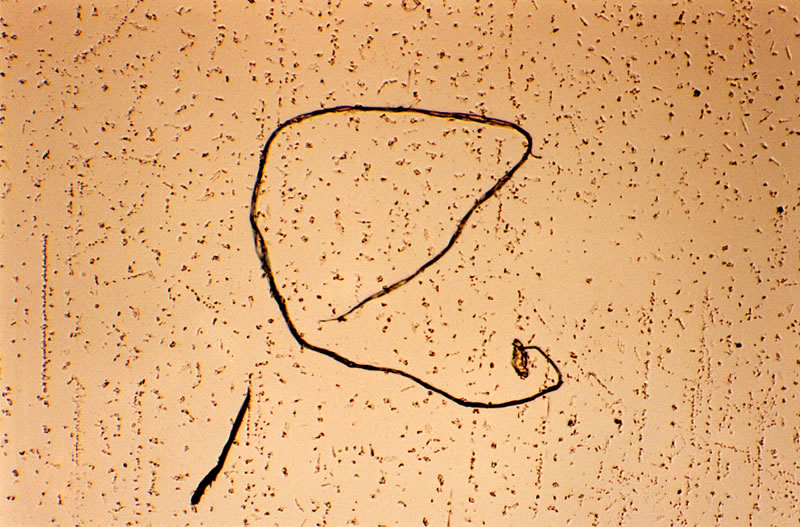
    A preparation from a patient’s brain who was diagnosed as alzheimer: see the nodules and a cellulose fibrille, which develop from uracil crystals.

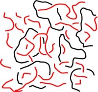
    Fibril and cellulose vesicles in a BSE preparation.

    This next image shows the ‘spikey’ formation that resembles exactly a fellow Morgellons sufferer has shown in microscopical pictures using blood and excreting yellowish liquid samples from his lesions.

    Blood samples taken from a patient who was diagnosed as cancer of bowels: see the prochlorons and hypha formations, which develop from these prochlorons.

    Reference: http://uracilandlife.org/uracilandlife_en.htm

    Lysosomes – Magnesium – Potassium – Calcium Beneficial for Morgellons Sufferers?

    •September 15, 2009 • 4 Comments

    As our research has revealed we believe that this insect virus, the Baculovirus,  is the causative agent of Morgellons Disease.

    We are stating that the Baculovirus is capable to ‘dock’ on to human cells.

    Research on this subject has shown that high Magnesium and high Potassium levels are the ‘key’ in eliminating the virus respectively inhibiting further re-production.

    My own experience in using these supplements, also adding higher doses of zinc, has lead me to the strong belief that these were beneficial in treating this disease as my symptoms have decreased tremendously.

    Here are a few excerpts that will show how I came to the above conclusions:

    “The Baculovirus is able to enter human hepatoma cells.”

    “Autographa californica multiple nucleopolyhedrovirus (AcMNPV), a prototype member of the Baculoviridae family, has gained increasing interest as a potential vector candidate for mammalian gene delivery applications.”

    “The host specificity of baculovirus was long supposed to be restricted to arthropods until Volkman and Goldsmith (53) showed that the viruses were efficiently taken up by mammalian cells. Later, Hofmann and colleagues (22) reported that the recombinant baculoviruses were also able to deliver genes into human hepatocytes.”

    “Entry of baculovirus to mammalian cells has been thought to be similar to that found in insect cells. Cell surface molecule interactions with baculovirus during uptake in mammalian cells are unclear; however, the virus has been suggested to use rather widely distributed and heterogeneous cell surface motifs (14). The first evidence for use of the endosomal pathway during baculovirus entry was provided by transducing cells in the presence of chloroquine, bafilomycin A1, and ammonium chloride, which all strongly prevented viral transduction.”

    “The early events of a viral infection include attachment of the virion to the receptor followed by entry into the cell and subsequent release of the genome. Despite numerous efforts, the natural receptor for baculovirus has not yet been discovered. Because of the wide range of mammalian cells that are susceptible to baculovirus transduction, it is likely that the target molecule is a relatively universal cell surface component, possibly heparan sulfate or even a phospholipid.”

    In conclusion, these data indicate that baculovirus uses at least clathrin-mediated endocytosis as one of its entry pathways to HepG2 cells. However, baculovirus attachment to clathrin-coated pits seemed to be a relatively rare phenomenon, and therefore other internalization mechanisms could also coexist.”

    “Clathrin-mediated endocytosis is known to lead to early and late endosomes, and finally to lysosomes (35). So far, many different viruses have been shown to utilize this pathway for release of the viral capsid to the cytoplasm from the early or late endosomes in mammalian cells.”

    “The size difference between clathrin-coated vesicles (100 to 150 nm; (35) and baculoviruses (25 x 260 nm) is likely to restrict viral entry via clathrin-mediated endocytosis into mammalian cells.”

    “Interestingly, Bilello et al. (4) have suggested that baculovirus entry to primary hepatocytes may require contact with the basolateral surface, which contains numerous microvilli (2). The occlusion-derived baculovirus has also been shown to fuse with the microvillar membrane of columnar epithelial cells in the highly alkaline midgut environment at low temperatures (38). In this study, baculovirus seemed to induce formation of microvilli and resided primarily on those areas of the cell surface (Fig. 3D). The higher occurrence of microvilli-like structures in virus-treated cells than in control cells may support the use of the macropinocytosis route in baculovirus entry.”

    “In summary, our results suggest that baculovirus entry into human hepatoma cells is a relatively slow process, probably due to the large size of the virus. Electron microscopy analysis showed viruses at the following multiple cellular locations: at the cell surface, in clathrin-coated pits, in early endosomes, and in intracellular vesicles resembling macropinosomes. In confocal microscopy and nanogold preembedding electron microscopy, the baculovirus particles were shown to colocalize with early and late endosomal/lysosomal markers. These results suggest that baculovirus may enter mammalian cells by both clathrin-mediated endocytosis and macropinocytosis.”

    What are Lysosomes?

    “Lysosomes are organelles containing digestive enzymes (acid hydrolases). They are found in animal cells, while in plant cells the same roles are performed by the vacuole. They digest excess or worn-out organelles, food particles, and engulfed viruses or bacteria. The membrane surrounding a lysosome allows the digestive enzymes to work at the 4.5 pH they require. Lysosomes fuse with vacuoles and dispense their enzymes into the vacuoles, digesting their contents.”

    image

    Proteins in different cellular compartments and structures tagged with green fluorescent protein.

    “Lysosomes are used for the digestion of macromolecules from phagocytosis (ingestion of other dying cells or larger extracellular material, like foreign invading microbes), endocytosis (where receptor proteins are recycled from the cell surface), and autophagy (wherein old or unneeded organelles or proteins, or microbes that have invaded the cytoplasm are delivered to the lysosome). Autophagy may also lead to autophagic cell death, a form of programmed self-destruction, or autolysis, of the cell, which means that the cell is digesting itself.

    Other functions include digesting foreign bacteria (or other forms of waste) that invade a cell and helping repair damage to the plasma membrane by serving as a membrane patch, sealing the wound.”

    Lysosomes are digestive enzymes as stated above. I personally think that buffering the ph level is essential to keep the body functions intact in order to control the symptoms of Morgellons Disease.

    Magnesium,Potassium and Calcium

    “Magnesium, the second most abundant intracellular cation, is a catalytic and structural element of major significance in the physiology of the human organism. Necessary for the anatomical and functional integrity of various subcellular organelles, it participates in all the major metabolic pathways, i.e., those involving carbohydrates, proteins and lipids, as well as in redox reactions. It is involved in the regulation of ion levels, maintaining the potassium level in the cell and exercising on the metabolism of calcium and phosphorus vitamin D-like effects.

    Integral to processes of defense, magnesium exhibits a variety of effects: antistress, anti-allergic, anti-anaphylactic, anti-inflammatory, antiradiation. Magnesium plays a role in thermoregulation; it stimulates phagocytosis and the formation of antibodies, complement and elements of the properdin system.

    Present in many tissues, it is active in the physiology of many systems, not only neuromuscular, osteo-articulatory, and dental, but also respiratory, endocrine, reproductive, ocular, digestive, hepatic, pancreatic, renal, cardiovascular and hematologic.”

    “Magnesium also appears to protect lysosomes. Magnesium deficit in the rat, for example, is accompanied by a degranulation of mast cells that may be compared to the degranulation of polynuclear basophils in man. This is not the result of a degranulating factor in serum, but rather of a direct cellular effect of magnesium deficit, perhaps a reduction in the level of cAMP.”

    “Magnesium thus acts as a major cellular and subcellular stabilizing agent which is necessary for the stability of plasma membranes, for the integrity of mitochondria, lysosomes, polysomes, and chromosomes as well as for the integrity of the helix of DNA and of messenger RNA and of RNA complexes.”

    “The effects of magnesium on leucocytes — enhancing phagocytosis and the production of lymphocytes and their transformation into blast cells while moderating the inflammatory reaction (342, 349) — are less important in the analysis of the effects of magnesium deficit on the formed elements of the blood as a vascular risk factor than are its stabilizing effects on platelets and erythrocytes.

    Magnesium deficit, on the other hand, favors vascular disease by its effects on the metabolism of phosphorus and calcium in calcinosis, on the metabolism of proteins, carbohydrates and glycoproteins in alterations analogous to connective tissue changes due to aging and finally on the metabolism of lipids and lipoproteins by enhancing atherogenic dyslipidemias “

    “Magnesium deficiency produces a true “aging” of bone with a slowed turnover of bone cells and a reduced receptivity to the hormone-like D vitamins as well as to parathyroid hormone. Magnesium deficiency affects the teeth before affecting skeletal bones.”

    Many Morgellons Sufferers have reported to have problems with their teeth and gums. Interesting is also the fact that Morgellons Sufferers have reported to have an exceedingly ‘craving’ for milk or milk products.

    “Magnesium deficiency produces low magnesium levels in the extra-cellular compartment and a reduction of levels in the cell along with hyperpermeability of the cell membrane. This depolarisation finally causes a lowered level of cellular potassium and a calcium overload (increase of intracellular Ca), in conjunction with a lowering of phosphorus levels and an increase in intracellular Na+. The increased influx of calcium into the cell produces lower blood levels of calcium and the release of potassium from the cell raises blood levels of potassium. Moreover, if the deficiency is prolonged, the cellular calcium overload may cause calcinosis, due to mixed apatite crystals which combine Ca, P and Mg.”

    Magnesium Deficiency, Enzymes, Energy and Detox

    “Magnesium is needed by every cell in the body including those of the brain and is one of the most important minerals when considering supplementation because of its vital role in hundreds of enzyme systems and functions related to reactions in cell metabolism, as well as being essential for the synthesis of proteins, for the utilization of fats and carbohydrates.Magnesium is needed not only for the production of specific detoxification enzymes but is also important for energy production related to cell detoxification. A magnesium deficiency can affect virtually every system of the body.”

    “Persons only slightly deficient in magnesium become irritable, highly-strung, and sensitive to noise, hyper-excitable, apprehensive and belligerent. If the deficiency is more severe or prolonged, they may develop twitching, tremors, irregular pulse, insomnia, muscle weakness, jerkiness and leg and foot cramps. If magnesium is severely deficient, the brain is particularly affected. Clouded thinking, confusion, disorientation, marked depression and even the terrifying hallucinations of delirium tremens are largely brought on by a lack of this nutrient and remedied when magnesium is given. Because large amounts of calcium are lost in the urine when magnesium is undersupplied, the lack of this nutrient indirectly becomes responsible for much rampant tooth decay, poor bone development, osteoporosis and slow healing of broken bones and fractures. With vitamin B6 (pyridoxine), magnesium helps to reduce and dissolve calcium phosphate kidney stones.”

    Possible manifestations of magnesium deficiency include:

    • Low energy
    • Fatigue
    • Weakness
    • Confusion
    • Nervousness
    • Anxiousness
    • Irritability
    • Seizures (and tantrums)
    • Poor digestion
    • PMS and hormonal imbalances
    • Inability to sleep
    • Muscle tension, spasm and cramps
    • Calcification of organs
    • Weakening of the bones
    • Abnormal heart rhythm

    Many of the above noted symptoms of Magnesium deficiency are similar to the physical symptoms of Morgellons Disease.

    “Magnesium is a crucial factor in the natural self-cleansing and detoxification responses of the body. It stimulates the sodium potassium pump on the cell wall and this initiates the cleansing process in part because the sodium-potassium-ATPase pump regulates intracellular and extracellular potassium levels. Cell membranes contain a sodium/potassium ATPase, a protein that uses the energy of ATP to pump sodium ions out of the cell, and potassium ions into the cell. The pump works all of the time, like a bilge pump in a leaky boat, pumping K+ and Na+ in and out, respectively.”

    “Potassium regulation is of course crucial because potassium acts as a counter flow for sodium’s role in nerve transmission. The body must put a high priority on regulating the potassium of the blood serum and this becomes difficult when magnesium levels become deficient.”

    “A magnesium deficiency can cause the body to lose potassium and this our bodies cannot afford. Within the cell wall is a sodium pump to provide a high internal potassium and a low internal sodium. Magnesium and potassium inside the cell assist oxidation, and sodium and calcium outside the cell wall help transmit the energy produced.”

    “Magnesium in general is essential for the survival of our cells but takes on further importance in the age of toxicity where our bodies are being bombarded on a daily basis with heavy metals. Magnesium thus protects the brain from toxic effects of chemicals.”

    Glutathione synthetase requires γ-glutamyl cysteine, glycine, ATP, and magnesium ions to form glutathione.[vii] In magnesium deficiency, the enzyme y-glutamyl transpeptidase is lowered.[viii] Data demonstrates a direct action of glutathione both in vivo and in vitro to enhance intracellular magnesium and a clinical linkage between cellular magnesium, GSH/GSSG ratios, and tissue glucose metabolism.[ix] Magnesium deficiency causes glutathione loss.”

    “Magnesium deficiency (MgD) has been associated with production of reactive oxygen species, cytokines, and eicosanoids, as well as vascular compromise in vivo.”

    References:

    http://jvi.asm.org/

    http://jvi.asm.org/cgi/content/abstract/77/1/328

    http://jvi.asm.org/cgi/content/full/79/24/15452

    http://en.wikipedia.org/wiki/Lysosome

    http://www.mgwater.com/durex01.shtml

    http://www.advanced-supplements.com/magnesium.html

    http://www.magnesiumforlife.com/detox_chelation.shtml

    Formation of Actin Filaments in Mammalian Cells – Baculovirus Protein FALPE and p10

    •September 12, 2009 • Leave a Comment

    FALPE and p10 Insect virus protein embedded in the Baculovirus might cause the proliferating formation of Actin Protein Filaments in humans bodies..mainly Morgellons Sufferers.

    As we know, the so called ‘fibers’ are seen to be the main symptom of this disease. 100% have reported to have them in/on lesions or have them emerging from intact skin or hair follicles.

    I believe that the infectious Baculovirus leads to an over reaction of Actin Protein filament production in our organism due to influence of the Insect virus proteins FALPE and p10.

    Here are a few excerpts out of the explanation of Actin Protein Filaments:

    Actin is a globular, roughly 42-kDa protein found in all eukaryotic cells.

    Actin is the monomeric subunit of two types of filaments in cells: microfilaments, one of the three major components of the cytoskeleton, and thin filaments, part of the contractile apparatus in muscle cells. Thus, actin participates in many important cellular processes including muscle contraction, cell motility, cell division and cytokinesis, vesicle and organelle movement, cell signaling, and the establishment and maintenance of cell junctions and cell shape.

    Principal interactions of structural proteins are at cadherin-based adherens junction. Actin filaments are linked to α-actinin and to the membrane through vinculin. The head domain of vinculin associates to E-cadherin via α-, β-, and γ-catenins. The tail domain of vinculin binds to membrane lipids and to actin filaments.

    The protein actin is one of the most highly conserved throughout evolution because it interacts with a large number of other proteins.

    All non-spherical prokaryotes appear to possess genes such as MreB, which encode homologues of actin; these genes are required for the cell’s shape to be maintained. The plasmid-derived gene ParM encodes an actin-like protein whose polymerised form is dynamically unstable, and appears to partition the plasmid DNA into the daughter cells during cell division by a mechanism analogous to that employed by microtubules in eukaryotic mitosis.

    Actin has four main functions in cells :

    • To form the most dynamic one of the three subclasses of the cytoskeleton, which gives mechanical support to cells, and hardwires the cytoplasm with the surroundings to support signal transduction.
    • To allow cell motility (see Actoclampin molecular motors), including phagocytosis of bacteria by macrophages.
    • In muscle cells to be the scaffold on which myosin proteins generate force to support muscle contraction.
    • In non-muscle cells as a track for cargo transport myosins [non-conventional myosins] such as myosin V and VI. Non-conventional myosins transport cargo, such as vesicles and organelles, in a directed fashion, using ATP hydrolysis, at a rate much faster than diffusion. Myosin V walks towards the barbed end of actin filaments, while myosin VI walks toward the pointed end. Most actin filaments are arranged with the barbed end toward the cellular membrane and the pointed end toward the cellular interior. This arrangement allows myosin V to be an effective motor for export of cargos, and myosin VI to be an effective motor for import.

    Pathway of Baculovirus in mammalian cells

    We have studied the infection pathway of Autographa californica multinuclear polyhedrosis virus (baculovirus) in mammalian cells.

    By titration with a baculovirus containing a green fluorescent protein cassette, we found that several, but not all, mammalian cell types can be infected efficiently.

    We demonstrate for the first time that this baculovirus can infect nondividing mammalian cells, which implies that the baculovirus is able to transport its genome across the nuclear membrane of mammalian cells.

    Our data further show that the virus enters via endocytosis, followed by an acid-induced fusion event, which releases the nucleocapsid into the cytoplasm.

    Cytochalasin D strongly reduces the infection efficiency but not the delivery of nucleocapsids to the cytoplasm, suggesting involvement of actin filaments in cytoplasmic transport of the capsids.

    In one study, nucleocapsids of a related baculovirus species (Ploidia interpunctella granulosis virus) were observed docking at the nuclear pore of infected insect cells, at different stages of releasing their genome, but not inside the nucleus.

    This suggests a mechanism of DNA transport similar to HSV.

    We show here that baculovirus can indeed infect nondividing mammalian cells through a mechanism apparently identical to that found in insect cells.

    The mode of nuclear entry of the viral genome appears to be different from what is known of other large DNA viruses.

    Our data suggest that the cigar-shaped nucleocapsid (25 nm in diameter) is transported through the nuclear pore, together with the viral genome.


    References:

    http://en.wikipedia.org/wiki/Actin

    http://www.pubmedcentral.nih.gov/articlerender.fcgi?artid=113992

    Enterobacteria – White Pox – Symptom

    •September 8, 2009 • 4 Comments

    According to a poll a fellow member has started at a Morgellons Disease Forum, 9 out of 10 participants claim to have white spots/pox on their skin.

    Significant is, that many Morgellons Sufferers seem to have them on their forearms.

    Some have reported, that these spots occurred after visual lesions and assume they could possibly be scar forming.

    In some cases these white pox marks can also appear without any previous formations of wounds/sores.

    A few members suggest Vitilligo as possible cause due to the fact that they look like a dysfunction of skin pigmentation in lack of Melanin in those areas.

    According to my research I believe that a bacteria, namely Enterobacteria http://en.wikipedia.org/wiki/Enterobacteriaceae, is  the cause of this appearance of white pox formations on skin.

    I claim this to be an additional main symptom of Morgellons Disease.

    Here is an image:  image

    This bacteria is associated with the so called ‘White Pox Disease’ that affects not only humans but also marine organisms.

    http://www.artificialreefs.org/Corals/diseasesfiles/Common%20Identified%20Coral%20Diseases.htm

    Here is an image of the bacterium Serratia marcescens on marine organism     image

    Another interesting aspect is, that Enterobacterium leads to an infection in insects such as flies of the type Drosophilia (fruit flies) but also other insects such as the silk worm.

    Some information on Drosophilia:

    http://en.wikipedia.org/wiki/Drosophila

    image

    I found it quite interesting that this bacterium is also commonly found in damp, moist environments such as bathrooms, toilets etc.,where it manifests as a pink water coloration and a slimy film. A few members have reported to have seen this in their facilities..

    Another aspect, I’ve found suspicious is the fact that this bacterium also can cause a biofilm on gums and teeth. Many Morgellons Sufferers have reported this condition.

    This article also states that Enterobacteria can occur as a manifestation of pink dis-coloration also in/on starch/sugar based foods.

    Potatoes for example are rich on starch and sugar.

    Research on the recent investigated fungus gnat has shown that these insects are attracted to this vegetable especially when in the decomposition stage.

    Many members of our forum have reported to have an environmental issue with fungus gnats, some have found these gnats harboring in/on potatoes or near where potatoes were stored.

    This investigation lead to the presumption that infections of Serratia marcescens (Enterobacteria) over an insect vector, such as the fungus gnat, is the main cause not only of our disease but is also suspected respectively to be highly related in causing those white pox/spots on our skin.

    Water contamination with this bacterium is also an important factor which I believe to be highly involved in causing Morgellons Disease.

    Unfortunately Enterobacteriae is multi resistant to several antibiotics and therefore very difficult to eradicate.

    This claims to be true, as many Morgellons Sufferers still remain ill after long term antibiotic treatments.

    For further information, here is the link explaining Serratia marcescens:

    http://en.wikipedia.org/wiki/Serratia_marcescens

    Excerpt:

    “Serratia marcescens is a species of Gram-negative, rod-shaped bacterium in the family Enterobacteriaceae.

    Due to its ubiquitous presence in the environment, and its preference for damp conditions, S. marcescens is commonly found growing in bathrooms (especially on tile grout, shower corners, toilet water line, and basin), where it manifests as a pink discoloration and slimy film feeding off phosphorous containing materials or fatty substances (such as soap and shampoo residue).

    S. marcescens may also be found in environments such as dirt, supposedly "sterile" places, and the subgingival biofilm of teeth.

    S. marcescens can cause infection in several sites, including the urinary tract, respiratory tract, wounds and the eye, where it may cause conjunctivitis, keratitis, endophthalmitis, and tear duct infections. It is also a rare cause of endocarditis and osteomyelitis (particularly in people who use intravenous drugs recreationally), pneumonia, and meningitis. Most S. marcescens strains are resistant to several antibiotics because of the presence of R-factors, which are a type of plasmid that carry one or more genes that encode resistance; all are considered intrinsically resistant to ampicillin, macrolides, and first-generation cephalosporins (such as cefalexin).

    In elkhorn coral, S. marcescens is the cause of the disease known as white pox disease

    In silkworms, it sometimes occurs as a secondary pathogen in viral flacherie disease.

    In Drosophila research laboratories, infection with S. marcescens is common.

    It manifests itself as a pink discoloration or plaque in or on larvae, pupae, or the usually starch and sugar-based food (especially when improperly prepared).

    Since 1950, S. marcescens has steadily increased as a cause of human infection, with many strains resistant to multiple antibiotics.

    The first indications of problems with the influenza vaccine produced by Chiron Corporation in 2004 involved S. marcescens contamination.

    Because of its red pigmentation, caused by expression of the pigment prodigiosin and its ability to grow on bread.”

    Serratia marcescens is also found as red pigmentation on bread and was used in influenza vaccines.

    Further research resulted that Enterobacteria can also infect Poultry and is also seen as major cause of disease in humans.

    http://www.vef.hr/org/bolesti_peradi/new_opportunities.htm

    Here an image of Enterobacteria in poultry:

    http://www.vef.hr/org/bolesti_peradi/bacilus%20cereus.jpg